Lesson type
Difficulty level

In this lesson, the simulation of a virtual epileptic patient is presented as an example of advanced brain simulation as a translational approach to deliver improved clinical results. You will learn about the fundamentals of epilepsy, as well as the concepts underlying epilepsy simulation. By using an iPython notebook, the detailed process of this approach is explained step by step. In the end, you are able to perform simple epilepsy simulations your own.
Difficulty level: Beginner
Duration: 1:28:53
Speaker: : Julie Courtiol

Course:
In this lesson you will learn how to simulate seizure events and epilepsy in The Virtual Brain. We will look at the paper On the Nature of Seizure Dynamics, which describes a new local model called the Epileptor, and apply this same model in The Virtual Brain. This is part 1 of 2 in a series explaining how to use the Epileptor. In this part, we focus on setting up the parameters.
Difficulty level: Beginner
Duration: 4:44
Speaker: : Paul Triebkorn

Manipulate the default connectome provided with TVB to see how structural lesions effect brain dynamics. In this hands-on session you will insert lesions into the connectome within the TVB graphical user interface (GUI). Afterwards, the modified connectome will be used for simulations and the resulting activity will be analysed using functional connectivity.
Difficulty level: Beginner
Duration: 31:22
Speaker: : Paul Triebkorn

This talk highlights a set of platform technologies, software, and data collections that close and shorten the feedback cycle in research.
Difficulty level: Beginner
Duration: 57:52
Speaker: : Satrajit Ghosh

Course:
This lesson provides an overview of the database of Genotypes and Phenotypes (dbGaP), which was developed to archive and distribute the data and results from studies that have investigated the interaction of genotype and phenotype in humans.
Difficulty level: Beginner
Duration: 48:22
Speaker: : Michael Feolo

Course:
Overview of the content for Day 1 of this course.
Difficulty level: Beginner
Duration: 00:01:59
Speaker: : Tristan Shuman

Course:
Best practices: the tips and tricks on how to get your Miniscope to work and how to get your experiments off the ground.
Difficulty level: Beginner
Duration: 00:53:34
Course:
This talk delves into challenges and opportunities of Miniscope design, seeking the optimal balance between scale and function.
Difficulty level: Beginner
Duration: 00:21:51
Speaker: : Susie Feng, Zach Pennington, Tycho Hoogland

Course:
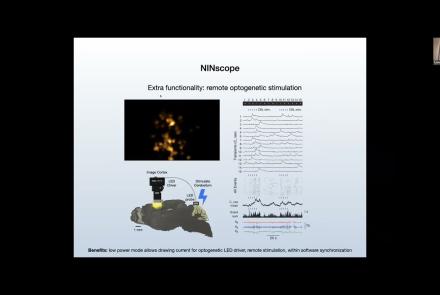
Attendees of this talk will learn aobut computational imaging systems and associated pipelines, as well as open-source software solutions supporting miniscope use.
Difficulty level: Beginner
Duration: 00:17:56
Speaker: : Susie Feng, Zach Pennington, Laura Waller

Course:
This talk covers the present state and future directions of calcium imaging data analysis, particularly in the context of one-photon vs two-photon approaches.
Difficulty level: Beginner
Duration: 00:21:06

Course:
In this talk, results from rodent experimentation using in vivo imaging are presented, demonstrating how the monitoring of neural ensembles may reveal patterns of learning during spatial tasks.
Difficulty level: Beginner
Duration: 00:19:43
Speaker: : Susie Feng, Zach Pennington, William Mau

Course:
How to start processing the raw imaging data generated with a Miniscope, including developing a usable pipeline and demoing the Minion pipeline.
Difficulty level: Beginner
Duration: 00:57:26
Speaker: : Daniel Aharoni, Phil Dong

Course:
The direction of miniature microscopes, including both MetaCell and other groups.
Difficulty level: Beginner
Duration: 00:49:16
Speaker: : Daniel Aharoni, Frederico N Sangiuliano

Course:
Overview of the content for Day 2 of this course.
Difficulty level: Beginner
Duration: 00:11:01
Speaker: : Tristan Shuman

Course:
Summary and closing remarks for this three-day course.
Difficulty level: Beginner
Duration: 00:04:56
Speaker: : Stephen Larson

This lecture covers infrared LED oblique illumination for studying neuronal circuits in in vitro block-preparations of the spinal cord and brain stem.
Difficulty level: Beginner
Duration: 25:16
Speaker: : Péter Szucs

This lecture covers the application of diffusion MRI for clinical and preclinical studies.
Difficulty level: Beginner
Duration: 33:10
Speaker: : Silvia de Santis

This talk covers the differences between applying HED annotation to fMRI datasets versus other neuroimaging practices, and also introduces an analysis pipeline using HED tags.
Difficulty level: Beginner
Duration: 22:52
Speaker: : Monique Denissen

This lesson provides a thorough description of neuroimaging development over time, both conceptually and technologically. You will learn about the fundamentals of imaging techniques such as MRI and PET, as well as how the resultant data may be used to generate novel data visualization schemas.
Difficulty level: Beginner
Duration: 1:43:57
Speaker: : Jack Van Horn

Course:
Longitudinal Online Research and Imaging System (LORIS) is a web-based data and project management software for neuroimaging research studies. It is an open source framework for storing and processing behavioural, clinical, neuroimaging and genetic data. LORIS also makes it easy to manage large datasets acquired over time in a longitudinal study, or at different locations in a large multi-site study.
Difficulty level: Beginner
Duration: 0:35
Speaker: : Samir Das